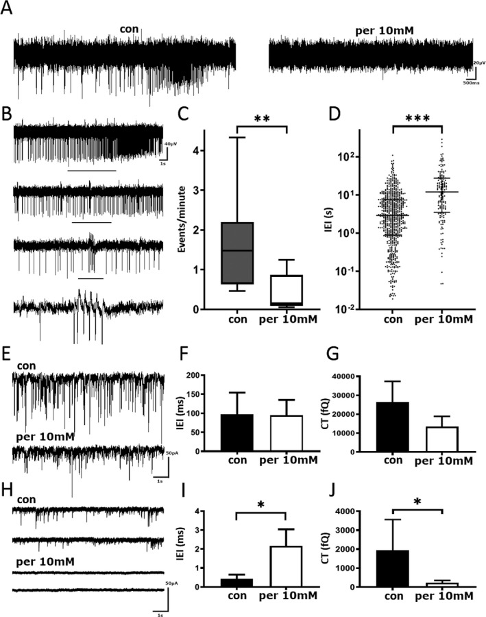
Figure 2.

Perampanel reduces spontaneous epileptiform activity in pediatric epilepsy tissue samples. (A) Example trace of local field potential recording before and after the addition of 10 µmol/L PER. Scale bar 20 µV versus 500 msec. (B) Example of spontaneous epileptiform activity. Black bar denotes zoomed in section of events. Scale bar 40 µV versus 1 sec. (C) Number of events per minute before and after the addition of 10 µmol/L PER. (D) Pooled interevent interval before and after application of 10 µmol/L PER. (E) Example trace of sIPSCs from pediatric human tissue before (top) and after (bottom) treatment with 10 µmol/L of Perampanel. Scale bar 50 pA versus 1 sec. (F) Pooled sIPSC inter‐event interval before and after application of 10 µmol/L PER (G) Pooled charge transfer of sIPSCs before and after application of 10 µmol/L PER. (H) Example trace of sEPSCs from pediatric human tissue before (top) and after (bottom) treatment with perampanel. Scale bar 50 pA versus 1 sec. (I) Pooled inter‐event interval before and after application of 10 µmol/L PER. (J) Pooled charge transfer before and after application of 10 µmol/L PER. PER, perampanel. *P ≤ 0.05; **P ≤ 0.01; ***P ≤ 0.001.
