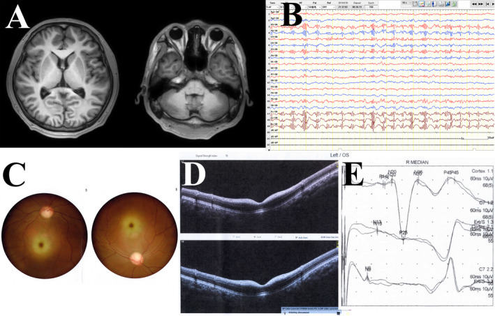
Figure 1.

Examination results of patient 1. (A) Axial T1 brain magnetic resonance imaging was normal; (B) electroencephalogram showed bilateral polyspike and polyspike‐slow waves in the central, parietal, and midline regions; (C) fundus examination showed bilateral cherry‐red spots; (D) optical coherence tomography indicated bilateral diffuse weakness of the retinal nerve fiber layer and ganglion cell complex, as well as the hyperreflexivity of the macular inner layer; (E) somatosensory evoked potential showed a huge potential of bilateral N20‐P25 in the upper limb pathway.
