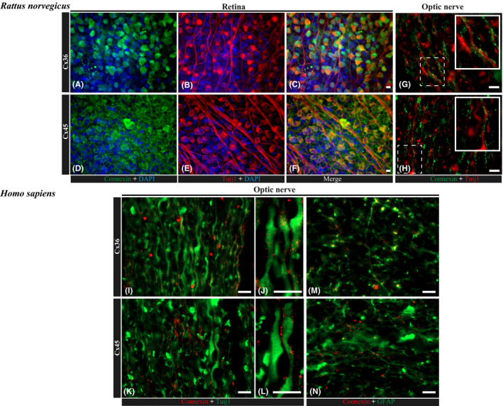
Figure 2.

Expression and localization of connexin 36 and 45 in rat and human retina and optic nerve. (A–F) immunofluorescence staining of rat, whole‐mounted retinas for Cx36 (A–C) and Cx45 (D–F) in colocalization with RGC marker, Tuij1 (β3tubulin); nuclei are counterstained with DAPI. Scale bar, 10 μm. In these whole‐mounted retina photographs, Cx36 expression within RGCs was present mostly intracellularly, while Cx45 showed a cell membrane pattern. In ON longitudinal sections, staining for both markers (Cx36 and Cx45) was observed along Tuij1‐positive axons. (G, H) immunofluorescence staining of paraffin longitudinal sections of a rat optic nerve head region for Cx36 (G) and Cx45 (H) in colocalization with RGC marker, Tuij1 (β3tubulin). Scale bar, 10 μm. (I–N) immunofluorescence staining of longitudinal paraffin sections of a human optic nerve head region for Cx36 (I, J) and Cx45 (K, L) in colocalization with RGC marker, Tuij1 (β3tubulin), and Cx36 (N) and Cx45 (M) in colocalization with glial cell marker, GFAP. Scale bars, 10 μm (I, K, N, M); 2 μm (J, L).
