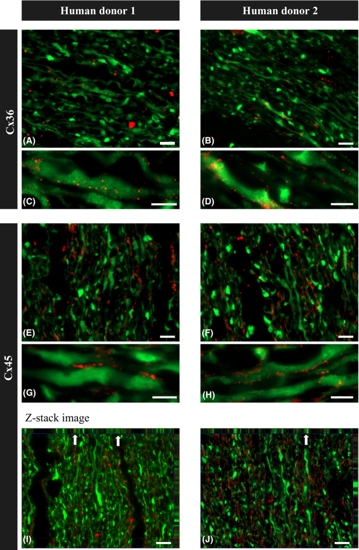
Figure 3.

Immunofluorescence staining of paraffin longitudinal sections of the ON from two healthy human donors. (A–D) immunostaining for Cx36 and Tuij1 showed dots of Cx36 staining colocalized with Tuij1‐positive ON axons. (E–H) immunostaining for Cx45 and Tuij1 showed dots of Cx45 staining colocalized with Tuij1‐positive ON axons. (I, J) photographs show Z‐stack imaging of longitudinal paraffin sections of the human optic nerve head region stained for Cx45 and Tuij1, with arrows pointing to the colocalization of axons with Cx45 projected in two planes. Scale bars, 10 μm (A, B, E, F, I, J); 5 μm (C, D, G, H).
