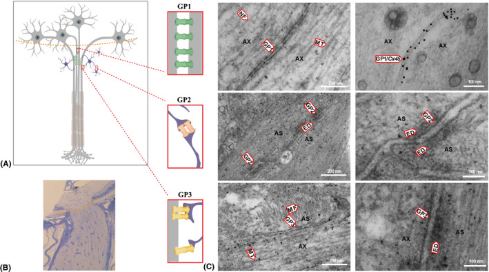
Figure 4.

Electron micrographs (EM) of longitudinal sections of a rat optic nerve head (ONH). (A) schematic picture depicting interconnections of axons and astrocytes within the ONH. There are three types of gap junctions identified in the ONH: gap junctions between two axons (GP1), between two astrocytes (GP2) and between axons and astrocytes (GP3). (B) light micrograph of a longitudinal section of rat optic nerve with marked level of ONH (red dashed line). (C) EM presenting different types of synaptic interconnections within the ONH. Upper panel shows fragments of two axons (AX) containing microtubules (MT), neurofilaments (NF) and gap junctions between two axons (GP1); the right EM shows GP1 with immunogold labelling of Cx45 within the gap junctions between two axons (GP1/Cx45). Middle panel shows an EM of fragments of two astrocytes (AS) showing gap junctions (GP2) between them. GP2 contains a double membrane and electron‐dense material (ED) around the electric synapse. The lower panel shows an EM of a fragment of an astrocyte (AS) and axon (AX) with a gap junction between them (GP3), microtubules (MT) in the axon, actin filaments (MF) in the astrocyte and electron‐dense material (ED) on the astrocytic side of GP3.
