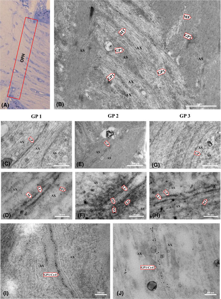
Figure 5.

Electron micrographs (EM) showing the ultrastructure of different interconnections within the rat optic nerve head. (A) light microscopy of the optic nerve head showing the region of EM imaging. (B) EM of a longitudinal ultrathin section of a rat optic nerve head showing the ultrastructure of cellular components: microtubules (MT) of axons (AX), microfilaments (MF) of astrocytes (AS) and gap junctions between two axons (GP1), between two astrocytes (GP2), and between axons and astrocytes (GP3). (C, D) EM showing a fragment of two axons with microtubule (MT) and GP1 details: lumen (LU), membrane (M). (E, F) EM of a fragment of two astrocytes and GP2 showing lumen (LU), inner membrane (IM), outer membrane (OM) and electron‐dense material (ED) around the gap junction. (G, H) EM of a fragment of the astrocyte and axon showing GP3 between them and microtubules in the axon. (I, J) immunogold labelling for Cx45; the EM shows a fragment of two axons with GP1 between them positively identified as composed of neuronal Cx45.
