
Figure 5.
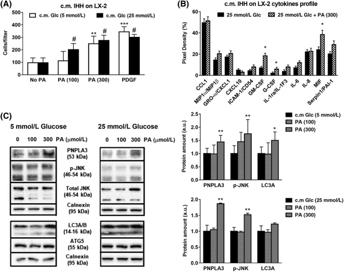
Conditioned medium (c.m.) collected from hepatocytes cultivated with PA and high glucose increases migration, pro‐fibrogenic and autophagy markers and cytokines release in HSCs. IHH cells cultivated with low (white bars) or high Glc (black bars) alone or in combination with PA (100 and 300 μmol/L). After 24 h, conditioned medium (c.m.) was collected and used to stimulate HSCs for 24 h. Conditions used in IHH cells are described in Material and Methods. A, Migration assay was performed in LX‐2 using Boyden chambers, as described above. PDGF (10 ng/ml) was used as positive control. **P < .01 and ***P < .001 vs c.m. No PA, 5 mmol/L Glc (white bar), #P < .01 vs No PA, 25 mmol/L Glc (black bar). B, Cytokine array quantification plot displayed and performed in media collected from LX‐2 previously exposed to IHH c.m., as described in Materials and Methods. *P < .05 vs 25 mmol/L Glc (black bars). C, Depicted expression of PNPLA3, phospho‐JNK (p‐JNK), total JNK, LC3A/B and ATG5 analyzed by western blotting. Calnexin used as loading control (protein amount reported as arbitrary units = a.u.). To evaluate pathway activity, p‐JNK measured in comparison with total JNK. Representative blot displayed. Densitometry analysis was calculated using ImageJ software. *P < .05, **P < .01 vs c.m. 5 or 25 mmol/L Glc (black bars)