
Figure 6.
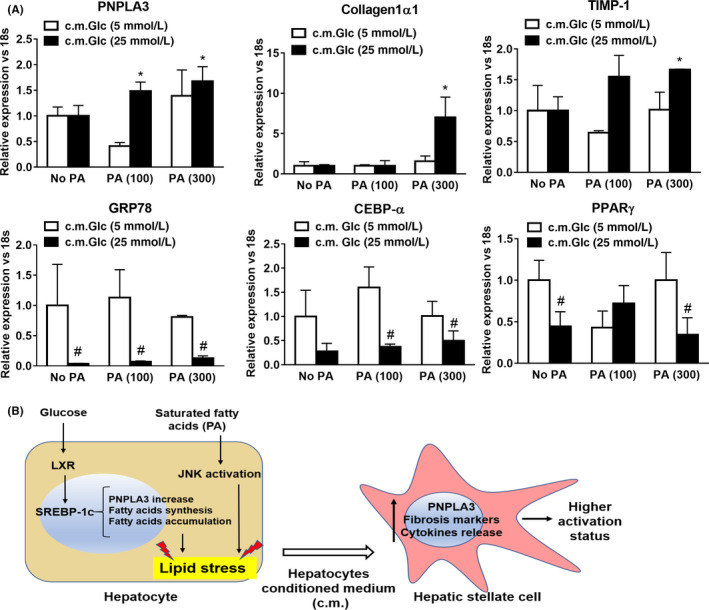
Glucose and Palmitic Acid trigger metabolic stress in hepatocytes, which in turn stimulates increase of PNPLA3, along with autophagic and pro‐fibrotic gene expression and inflammatory cytokine secretion in HSCs. IHH cells cultivated with low (white bars) or high Glc (black bars) alone or in combination with PA (100 and 300 μmol/L). After 24 h, conditioned medium (c.m.) was collected and used to stimulate HSCs for 24 h. Conditions used in IHH cells are described in Material and Methods. A, Expression of PNPLA3, Collagen1α1, TIMP‐1, GRP78, CEBPα and PPARγ analyzed by real‐time PCR and normalized to 18 s. *P < .05 vs c.m. No PA, 25 mmol/L Glc (black bars). #P < .01 vs 5 mmol/L Glc (white bars) relative to the same PA concentration. B, Representative scheme resembling PNPLA3 and de‐novo lipogenesis induction through SREBP‐1c and LXR by Glc (25 mmol/L). In addition, saturated FFA such as PA, induce lipid accumulation and cellular stress. Exposure of HSCs to c.m. from lipid stressed hepatocytes results in PNPLA3 increase and aggravates pro‐fibrogenic features (migration, Collagen1α1, α‐SMA, TIMP‐1, cytokine release and autophagic markers) in HSCs