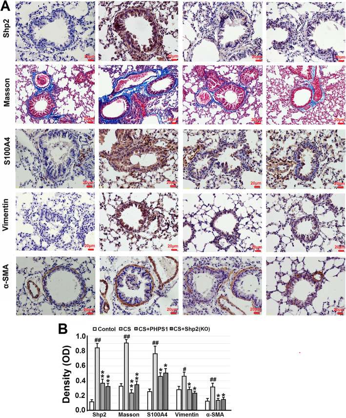
Fig. 3.
Genetic ablation of Shp2 in lung epithelial cells or pharmacological inhibition of Shp2 ameliorates collagen deposition and the expression of EMT-related proteins in lung tissues from the CS-exposed mice. a Representative images show that Shp2 knockout in lung epithelial cells or Shp2 inhibitor – PHPS1 (3 mg/kg, daily i.p injection) significantly reverses the CS-induced collagen deposition (Masson’s trichrome staining – blue color) and immunohistochemical signal (brown color) of Shp2, S100A4, vimentin and α-SMA in lung tissue sections. b The optical density value of Masson’s trichrome staining and immunohistochemical staining are quantified. Data are presented as the mean ± SEM. n = 8 per group. Scale bar = 20 μm. #p < 0.05, ##p < 0.01, compared with control (WT mice), *p < 0.05, **p < 0.01 compared with control (WT mice) with CS exposure, one-way ANOVA followed by the Student-Newman-Keuls test

