Fig. 2.
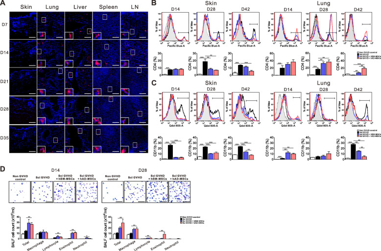
Migration of hMSCs into each organ and analysis of immune cell infiltration into skin and lung tissues. a PKH-26-labeled hAD-MSCs were injected into the tail vein on 3, 5, and 7 days after allo-HSCT. The skin, lung, liver, spleen, and lymph node (LN) sections were analyzed by confocal microscopy for the detection of PKH-26-positive cells (shown in red). Flow cytometric analyses were performed using skin and bronchoalveolar lavage fluid (BALF). b, c Frequency of CD4 T cells (b) and CD11b (c) cells 14, 28, and 42 days after transplantation. d Total BALF cell count 14 and 28 days after transplantation. Each value indicates the mean ± SEM of 4–9 mice per group. *P < 0.05; **P < 0.01, ***P < 0.001

