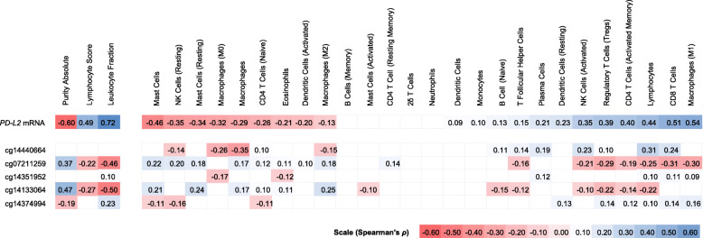
Fig. 3.
Heatmap of association between PD-L2 CpG site methylation and mRNA expression with tumor-infiltrating lymphocytes (TILs) according to Thorsson et al. [27]. Shown are Spearman’s rank correlations (Spearman’s ρ) between methylation / mRNA expression of PD-L2 and leukocyte fraction, as well as tumor-infiltrating leukocytes, including lymphocytes (CD8+ T cells, regulatory T cells, γδ T cells, naïve CD4+ T cells, resting and activated memory CD4+ T cells, naïve B cells, memory B cells, and resting and activated natural killer cells), monocytes and macrophages (M0 / M1 / M2 macrophages), resting and activated dendritic cells, resting and activated mast cells, eosinophils, and neutrophils. Immune signatures of tumor-infiltrating leukocytes were based on RNA-Seq analysis and the leukocyte fraction was based on methylation analysis. Only statistically significant (P < 0.05) are shown in color

