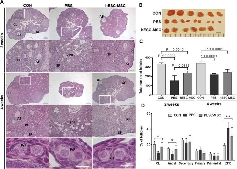
Fig. 4.
Recovery of ovarian structure by hESC-MSC transplantation in cisplatin-induced ovarian failure. a Ovarian histology was analyzed 2 and 4 weeks after transplantation using H&E staining. Scale bar = 200 or 50 μm. PrFs in insets were captured from other section. b Ovaries were removed from mice in the control, cisplatin + PBS, and cisplatin + hESC-MSC transplantation groups. Scale bar = 2 mm. c Total number of follicles per ovary. d Percentages of each follicle type per ovary. *P < 0.05, **P < 0.001; CL, corpus luteum; AF, antral follicle; SF, secondary follicle; PF, primary follicle; PrF, primordial follicle; and ZPR, zona pellucida remnant

