
Fig. 6.
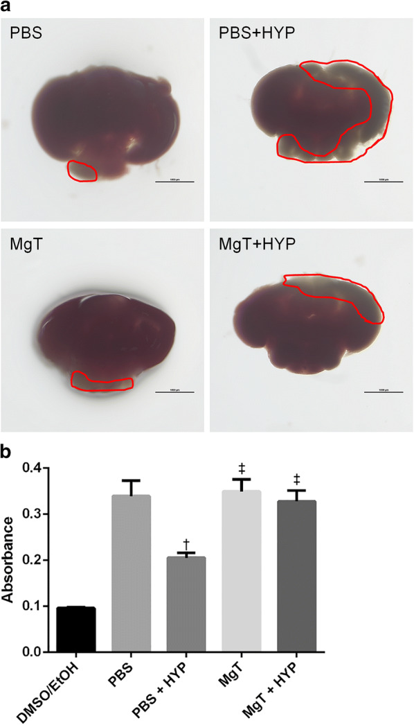
Zebrafish brain injury detected by TTC staining. a TTC-stained zebrafish brain sections: PBS, PBS + HYP, MgT, and MgT + HYP. Scale bar = 1000 um. The square indicates an unstained area. bSpectrophotometric measurement. Compared to the PBS group, the PBS + HYP group showed significantly low absorbance, while the MgT or MgT + HYP group did not. Eight brains in each group were analyzed. Data are shown as mean ± SEM. †p < 0.05 compared to the PBS group, ‡p < 0.05 compared to the PBS + HYP group. PBS, phosphate-buffered saline; MgT, magnesium l-threonate; HYP, hypoxia. TTC, 2,3,5-triphenyltetrazolium chloride