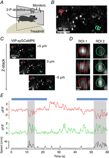
Figure 2. Imaging synaptic activity in awake behaving mice.

A, basic experimental set up for in vivo imaging. Awake animals are head‐fixed under two‐photon (2‐P) excitation while the animal is allowed to run freely on a polystyrene treadmill connected to a rotary encoder to record running speed. Visual stimuli are delivered through two computer monitors placed in front of the animal. B, example two‐photon image of a small field of synapses labelled with SyGCaMP6f. C, two‐photon images of the same field of synapses at imaging depths separated by 5 µm intervals. D, the synapses circled in B and C exhibit significant changes in intensity when the focal plane changes over a few microns. E, uncorrected fluorescence time‐series compared to locomotor behaviour for the same synapses. Blue bars show the timing of a stimulus (full‐field drifting grating).
