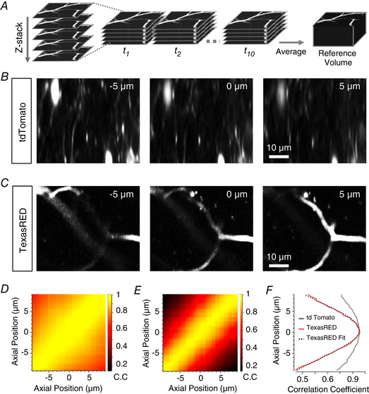
Figure 4. An anatomical marker of z‐position: a comparison of tdTomato and dextran‐TexasRED in blood vessels.

A, schematic showing construction of reference anatomical volume. Multiple stacks are rapidly acquired to enable registration of volumes before collapsing to an average volume. B, two‐photon images of VIP interneurons in mouse V1 labelled with tdTomato. Images are example slices from a reference stack acquired as in A at 5 µm above (left panel) or below (right panel) a central plane (central panel). C, as in B but with blood vessels labelled with dextran‐TexasRED. D and E, cross‐correlation matrices of an average reference stack with individual slices from a single registered volume for tdTomato (D) and labelled blood vessels (E). F, cross‐correlogram of the central slice of the volume with the reference stack for tdTomato (grey) and labelled blood vessels (red). A Gaussian (dotted line) is fitted to the TexasRED correlogram (= 8.9 µm) and the maximum taken as the estimated axial location of this slice. Note that the modulation of the correlation coefficient as a function of displacement is much larger for capillaries filled with TexasRED compared to the neurons labelled with tdTomato.
