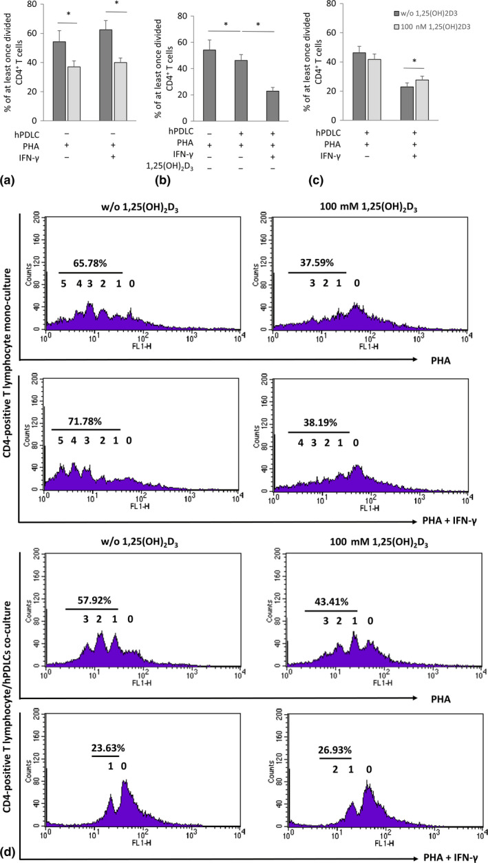
Figure 1.

Effect of 1,25(OH)2D3 on CD4+ T lymphocytes proliferation in the presence and absence of hPDLCs. Allogenic CD4+ T lymphocytes were activated by 10 µg/ml PHA and co‐cultured with 100 ng/ml IFN‐γ and 100 nM 1,25(OH)2D3 stimulated hPDLCs for 5 days in an indirect co‐culture model (b, c). PHA‐activated CD4+ T lymphocytes, stimulated with different stimuli in the absence of hPDLCs, served as control (A). T‐lymphocyte proliferation was assessed by determining the percentage of at least once divided CFSE‐labelled CD4+ T lymphocytes using flow cytometry. (a‐c) show data as mean value ± SEM from five independent experiments with hPDLCs isolated from five different individuals. *p‐value < .05 compared between appropriate groups as indicated. (d) shows representative data of one CD4+ T‐lymphocyte proliferation assay experiment presented in a one‐parameter histogram. The percentage of at least once divided CD4+ T lymphocytes is given. 0 presents the parental generation. 1, 2, 3, 4, 5 and 6 present the first, second, third, fourth, fifth and sixth generation, respectively
