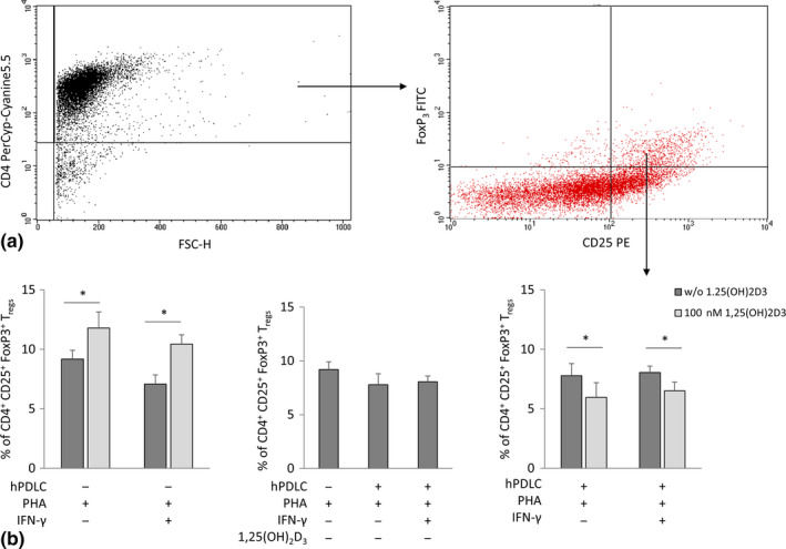
Figure 2.

Effect of 1,25(OH)2D3 on the percentage of CD4+ CD25+ FoxP3+ Tregs in the presence and absence of hPDLCs. Allogenic CD4+ T lymphocytes were activated by 10 µg/ml PHA and co‐cultured with 100 ng/ml IFN‐γ and 100 nM 1,25(OH)2D3 treated hPDLCs for 5 days in an indirect co‐culture model. PHA‐activated CD4+ T lymphocytes stimulated with different stimuli in the absence of hPDLCs served as control. CD4, CD25 and FoxP3 expression was estimated by immunostaining, followed by flow cytometry analysis. Representative dot plots show the gating strategy of flow cytometry analysis. After gating CD4+ T lymphocytes, FoxP3/CD25 double‐positive T lymphocytes were determined (a). Subsequently, the percentage of CD4+ CD25+ FoxP3+ Tregs were determined and presented as mean value ± SEM from five independent experiments with cells isolated from 5 different individuals (b). *p‐value < .05 compared between appropriate groups as indicated
