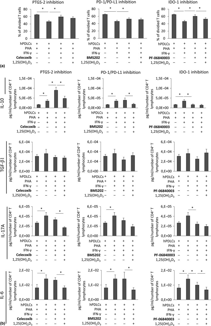
Figure 7.

Effect of IDO‐1, PD‐L1 or PTGS‐2 inhibitors on the proliferation and the production of IL‐10, TGF‐β1, IL‐17A and IL‐6 in CD4+ T lymphocytes in the presence of IFN‐γ and 1,25(OH)2D3 treated hPDLCs. Allogenic CD4+ T lymphocytes were activated by 10 µg/ml PHA and co‐cultured with IFN‐γ and 1,25(OH)2D3 stimulated hPDLCs for 5 days in an indirect co‐culture model. Additionally, either 50 µM IDO‐1 inhibitor PF‐06840003 or 1 µM PD‐1/PD‐L1 interaction inhibitor BMS202 or 1 µM PTGS‐2 inhibitor Celecoxib were added to appropriate hPDLCs before and during indirect co‐culture. CD4+ T‐lymphocyte proliferation was verified by determining the percentage of at least once divided CFSE‐labelled CD4+ T lymphocytes by flow cytometry (a). Additionally, IL‐10, TGF‐β1, IL‐17A and IL‐6 protein levels in conditioned media were determined by appropriate ELISA (b). All data are presented as mean value ± SEM from five independent experiments with hPDLCs isolated from five different individuals. * p‐value < .05 compared between groups as indicated
