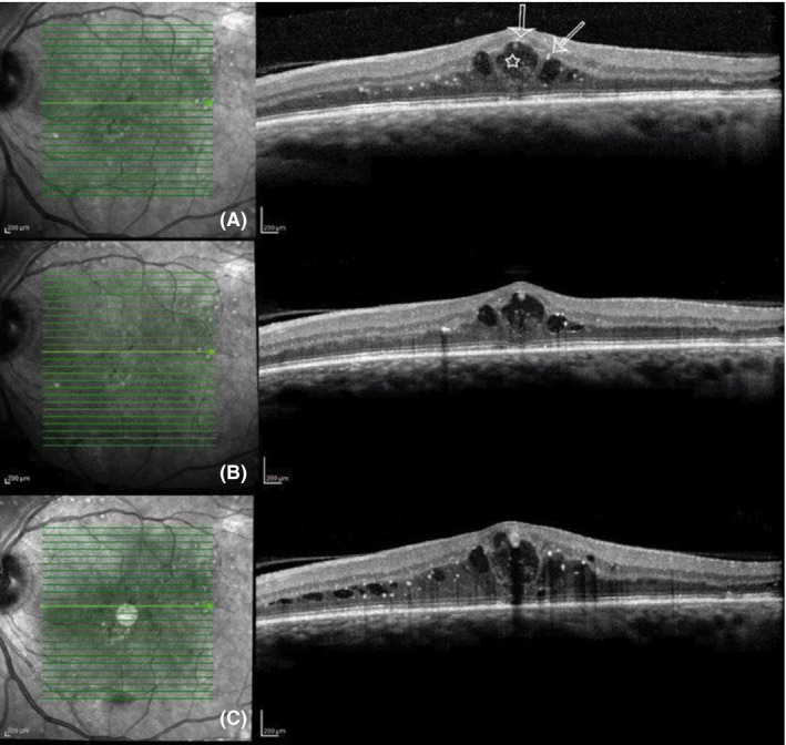
Figure 3.

Example of a patient treated with a dexamethasone implant with cytokine response at week 2 and missing morphological improvement. (A) Baseline optical coherence tomography (OCT) scan. The arrows mark the hyperreflective foci at the cyst borders. The star marks the intraretinal partly fibrotic tissue. (B) OCT at week 2. Cytokine levels decreased, but morphological and functional outcomes remain similar. (C) OCT at week 20. Cytokine levels increased after three months. Increased clinically significant macular oedema.
