
Figure 3.
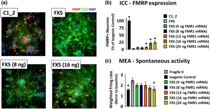
Transfection of FMR1 mRNA into FXS cultures results in a transient FMRP mosaic culture. (a) Immunofluorescent images from C1_2 neurons (upper left panel) and FXS neurons (upper right panel) in which 8ng FMR1 mRNA (lower left panel) and 16ng FMR1 mRNA (lower right panel) have been transiently transfected. Blue—nuclei, green—β‐Tubulin and red—FMRP. (b) Quantification of FMRP‐positive neurons in cultures 24 hr following transfection with varying amounts of FMR1 mRNA shows an increasing number of FMRP‐positive neurons with increasing amounts of FMR1 mRNA. Data normalized to C1_2 cultures. (c) Quantification of spontaneous activity, normalized to a pre‐transfection baseline, recorded on MEAs beginning 24hr post‐transfection of varying amounts of FMR1 mRNA, shows reduced neuronal activity in FXS neuronal cultures in which greater than 25% FMRP‐expressing neurons are present. Error bars represent ± SEM [Colour figure can be viewed at wileyonlinelibrary.com]