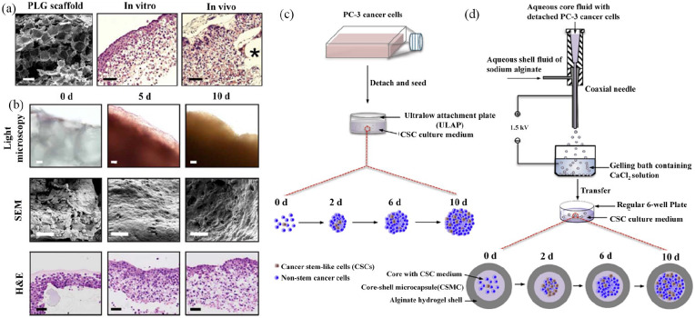
Figure 3.
3D cancer stem cell (CSC) models developed with porous scaffolds (a) and (b) and hydrogels (c) and (d). Characterization of tumor model (a) and (b): (a) Microstructure of cells loaded onto 3D PLG scaffolds visualized under scanning electron microscopy (SEM), and tumor-like tissue hematoxylin and eosin (H&E) stained specimens photomicrographs. Asterisk symbol (*) represent fragments of polymeric scaffolds. (b) Light microscopy, microstructure under SEM, and H&E-stained microphotographs of tumor-like tissues cultured in 3D PLG at different time points. Reproduced with permission from Fischbach et al.56 (2007, Nat Methods). Enrichment of PC-3 human prostate CSCs using conventional bulk suspension culture versus miniaturized 3D culture (c) and (d): (c) Schematic illustration of conventional bulk suspension culture in ultralow attachment plate. (d) Schematic illustration of miniaturized 3D culture in regular 6-well plates. The core of core-shell microcapsules (CSMCs) containing PC-3 prostate cells was prepared through coaxial electrospray by a coaxial needle. CSMCs were then cultured in regular 6-well plates for at least 10 days. Reproduced with permission from Rao et al.75 (2014, Biomaterials).

