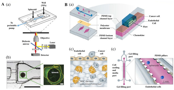
Figure 4.
3D cancer stem cell (CSC) models developed using microfluidic devices. (A, a, b) Schematic of the tumor-on-a-chip. (A, a) Schematic of the polydimethylsiloxane microfluidic device at the microscope stage. (A, b) The microfluidic device (left) showing the channel width of 600 mm at the inlet, which extends to 1200 mm in the imaging chamber where the spheroid is immobilized. The height of the channel is 250 mm and decreases to 25 mm at the end of the imaging chamber, forming a dam. A spheroid (right) stained for 10 min with anti-Laminin-FITC, then flushed for 5 min with imaging media. (B, a-c) Schematic of the organ-on-a-chip model. Reproduced with permission from Albanese et al.85 (2013, Nat Commun). (B, a) An endothelium-on-a-chip microvascular established in a segmented microfluidic channel allowed endothelial cells cultured on a chemokine supplemented permeable membrane to undergo activation and basal stimulation during the investigation of attached circulating cancer cells in breast tumor metastasis. The impacts of chemokines, including tumor necrosis factor, were examined via incorporating the chemokines in the bottom channel. The endothelium pre-treated with tumor necrosis factor attracted more tumor cells compared to the untreated endothelium. (B, b) Migration of breast tumor cells to the bone was examined in microfluidic device with endothelial cell culture from human umbilical vein next to bone cells derived from mesenchymal stem cells of the human bone marrow held a 3D collagen gel. Movement of cancerous cell to the bone was detected. (B, c) To assess the epithelial-mesenchymal transition (EMT) during malignancy, spheroids of lung cancer were fixed in micropatterned 3D matrices connected closely to endothelial cells linning on the microchannel. EMT examination was performed with microfluorometry to identify cancer spheroids distribution. Reproduced with permission from Esch et al.86 (2015, Nat Rev Drug Discov).

