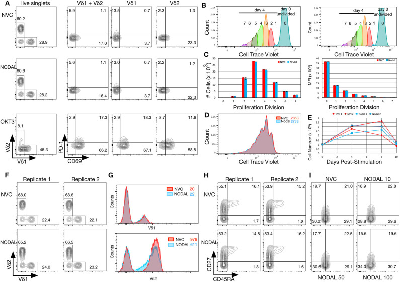
Figure 3.
NODAL stimulation has no impact on activation markers, proliferation or maturation of γδ T cells but longer term exposure to NODAL results in decreased Vδ2 TCR surface expression. (A), γδ T cells were subject to 24 h 100 ng/ml NODAL stimulation, and then surface expression of Vδ1 and Vδ2 TCR, PD-1, and CD69 were assessed via flow cytometry. NVC, NODAL vehicle control; anti-CD3 (OKT3) stimulation was done as a positive control for activation markers. This is a representative example of n = 3 independent experiments. (B), Long term NODAL stimulation has no impact on γδ T cell proliferation. γδ T cells from the same culture as in A were labeled with Cell Trace Violet, then stimulated one time with NODAL vehicle control (NVC) or 100 ng/ml recombinant human NODAL. Flow cytometry was performed on days 0, 4, 8, and 10. Proliferation modeling analyses for the day 4 time point are shown. (C), Cell numbers and median fluorescence intensities (MFI) of proliferation divisions for the experiment shown in B are shown; n = 2 technical replicates, error bars are SD. (D), A representative example of histogram overlays from the day 4 time point of the experiment shown in B and C; MFIs are indicated. (E), Cell counts on days 0, 4, 8, and 10 are plotted here for the two technical replicates done in the experiment shown in (B–D). No significant differences were observed. (F), Ten days after NODAL stimulation, cells cultured in parallel with the experiment in B-E were stained for Vδ1 and Vδ2 TCR expression; results from two technical replicates are shown. (G), Histogram overlays and MFIs of TCR expression for representative examples from the experiment shown in (F). (H), For the experiment shown in (F,G), maturation was assessed via CD45RA and CD27 staining; both technical replicates are shown. (I), Maturation assessment after stimulating γδ T cells for 4 days with 10, 50, or 100 ng/ml recombinant human NODAL.

