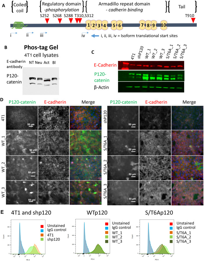
Fig 1. Establishment of p120-catenin phosphorylation dead mutant 4T1 cell lines.
(A) Schematic showing the location of the 6 mutated phosphorylation sites in p120-catenin, indicated by red triangles. 5 of the ser/thr sites are located in the N-terminal putative regulatory domain, and 1 is in the C-terminal tail domain. (B) Decrease in p120-catenin phosphorylation observed in 4T1 cells when treated with different E-cadherin functional antibodies (3μg/ml) using a phos-tag gel. (NT: No Treatment, Neu: Neutral Antibody, Act: Activating Antibody, Bl: Blocking Antibody) (C) Expression of p120-catenin and E-cadherin protein levels by western blot in 6S/T>A phospho-mutants (S/T6A_1, S/T6A_2, S/T6A_3) or Wildtype p120-catenin isoform 3 (WT_1, WT_2, WT_3) expressing 4T1 cell lines established from single cell clones after knockdown of endogenous p120-catenin with shRNA (shP120). (D) Immunofluorescent staining of p120-catenin and E-cadherin in the 4T1 clonal cell lines established. (E) Flow cytometry staining showing rescue of surface E-cadherin levels of expression after re-expression of either WT or S/T6A p120-catenin.

