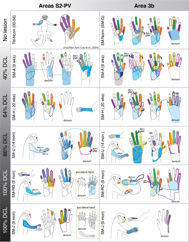
Figure 1.
Schematic drawings of representative neuronal receptive fields determined from microelectrode recordings in areas 3b and S2/PV. The location and size of receptive fields are color-coded and outlined on drawings of the face, body, and glabrous and dorsal surfaces of the hand. Note that the purpose of this illustration is to depict overall differences in receptive field locations and sizes across monkeys with different dorsal column lesion (DCL) extents and recovery times. Abbreviations: dor+glab, dorsal hair/skin and glabrous skin; dor+v, dorsal and ventral (hair/skin).

