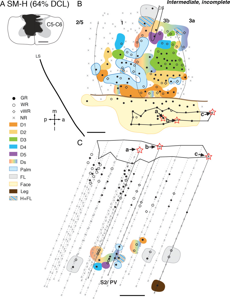
Figure 2.
Representative case for Group 1. Somatotopic maps of areas 3b, 1, and secondary somatosensory cortex (S2)/parietal ventral (PV) of squirrel monkey SM-H after an intermediate-term, incomplete DCL (64% complete; 142 days) at the C5–C6 level. A. Drawing shows the reconstructed transverse view of DCL in the spinal cord. Black shading depicts the area of tissue loss, dark gray surrounding the black shading depicts the area with abnormal tissue, and the light gray depicts the gray matter of the spinal cord. B. The reconstructed somatotopic map shows that neurons in hand region of area 3b responded well (shown in solid circle) with a nearly normal somatotopy. The hand region in area 1 is slightly less responsive. C. The reconstructed somatotopic map of the hand region in areas S2/PV obtained from deep penetrations. Responses to touch on single digit, multiple digits, or larger areas involving the arm and hand were found between the representations of arm and shoulders that are located rostrally (presumably PV) and caudally (presumably S2). Each symbol (i.e., solid circle, circle, diamond, and x) depicts the location and neuronal responsiveness of one mapping site. Red stars mark the locations of electrolytic lesions made along those electrode penetrations. Neuronal receptive fields in the body parts are color-coded, and color-coded stripes mark combinations of receptive fields in different body parts. Abbreviations: 3a, 3b, 1, 2/5, S2/PV, areas 3a, 3b, 1, 2/5, and S2/PV; a, anterior; C5–C6, cervical segments of C5–C6; CS, central sulcus; D1–D5, digits 1–5; Ds, multiple digits; FL, forelimb; GR, good response; H, hand; H + FL, hand and forelimb; l, lateral; LS, lateral sulcus; m, medial; NR, no response; p, posterior; vWR, very weak response to hard taps; WR, weak response; Scale bar is 1 mm in A, B, C.

