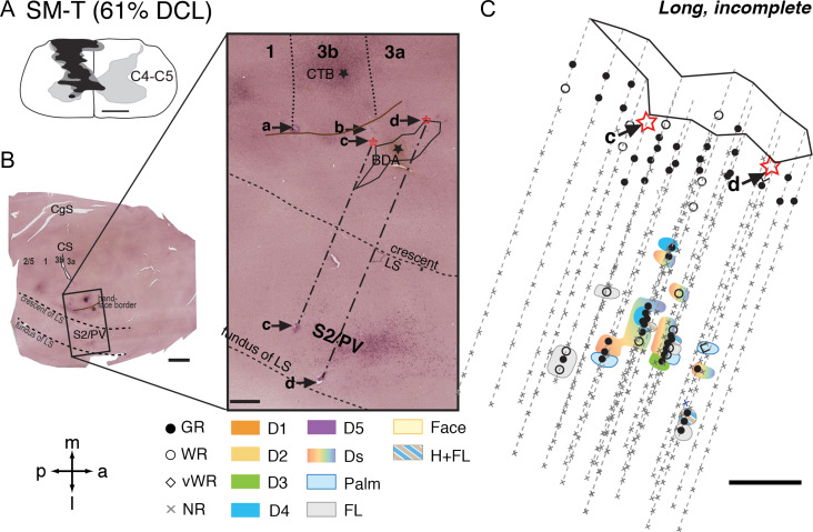
Figure 3.
Representative case for Group 2. Alignment of the somatotopic map of areas S2/PV and brain sections stained for cholera toxin subunit B (CTB) in monkey SM-T after a long-term, incomplete DCL (61% complete; 321 days) at the C4–C5 level. A. Drawing shows the reconstructed transverse view of DCL in the spinal cord. B. Photomicrographs of flattened cortical sections immunoreacted for CTB labeling showing CS, unfolded LS, and locations of strategically placed electrolytic lesions of a, b, c, and d. Among these, “c” and “d” are deep microelectrode penetrations inserted from the pia surface in the face region of area 3b into the hand region in S2/PV in the upper bank of lateral sulcus. C. The reconstructed somatotopic map shows responses encountered from deep penetrations recorded in areas S2/PV. Scale bar is 5 mm in the low magnification image of panel B (left), and is 1 mm in all others. Abbreviations: C4–C5, cervical segments of C4–C5; CgS, cingulate sulcus. Other conventions follow Figure 2.

