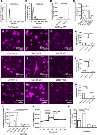
Fig. 2. Enhanced MCT1 and CD147 in hypoxic U251 GMs promote intracellular Ca2+-dependent exosome release.

(A and B) Size distribution and quantity of exosomes released from normoxic and hypoxic GMs for 24 hours (NTA analysis). (C) Enhanced release of exosomes from hypoxic GMs (versus normoxic GMs). (D) Analysis of exosome release from GMs treated with control 1, MCT1 OE, MCT1 KD, CD147 OE (all lentivirus), and control 2 and CD147 KD (antisense oligonucleotides) constructs. (E to P) Representative images of Fura Red calcium dye- loaded- hypoxic (versus normoxic), MCT1 OE- or MCT1 KD- (versus control 1) induced, CD147 OE- or CD147 KD- (versus control 1 & 2) induced, and BAPTA-AM (20 μM)-treated GMs. (Q) Enhanced exosome release from MCT1 OE– and CD147 OE–induced (versus control 1) GMs, followed by a marked decline in exosome release by treatment with BAPTA-AM (20 μM, 100 μl). (R) Enhanced intracellular Ca2+ levels in GMs by treatment with sodium-l-lactate (20 mM, 100 μl), followed by distinctive decline in intracellular Ca2+ level by treatment with BAPTA-AM (20 μM, 100 μl). (S) NTA exosome release assay from GMs exposed to four different conditions for 10 min described in Materials and Methods. Briefly, a, Exo–fetal bovine serum (FBS) medium; b, sodium-l-lactate (20 mM), c, BAPTA-AM; d, BAPTA-AM with the pretreatment of sodium-l-lactate (20 mM). All chemicals were dissolved in the Exo-FBS medium containing 1% dimethyl sulfoxide. All data were shown as the means ± SD. Significance level: **P < 0.01, *P < 0.05, hypoxia versus normoxia, BAPTA-AM versus control, MCT1 KD lentivirus versus empty backbone lentivirus (control 1), and CD147 antisense oligonucleotides versus antisense control oligonucleotides (control 2).
