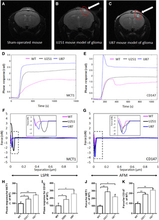
Fig. 6. Label-free sensitive detection of MCT1 and CD147 in serum-derived exosomes from a mouse model of glioma.

(A to C) Representative MRI images for the brain of sham-operated mice and U251 and U87 mouse models of glioma. (D and E) Representative phase responses of the LSPR biosensor with the functionalized SAM-AuNIs sensing chip with anti-MCT1 AB or anti-CD147 AB and (F and G) representative separation force curves of the AFM biosensor with the functionalized silicon nitride cantilever tip with anti-MCT1 AB or anti-CD147 AB toward serum-derived exosomes from sham-operated mice and U251 and U87 mouse models of glioma. (H to K) Bar graph summarizing the relative strength of LSPR responses (n = 3) or AFM forces (n = 3) toward exosomal MCT1 [e.g., (D) and (E)] and CD147 [e.g., (F) and (G)]. Detailed processes of LSPR and AFM biosensing were described in Materials and Methods. All data were expressed as the means ± SD. Significance level: **P < 0.01, *P < 0.05, U251 or U87 mouse model of glioma versus sham-operated severe combined immunodeficient mouse. WT, wild type.
