. 2020 Jun 26;9:e56945. doi: 10.7554/eLife.56945

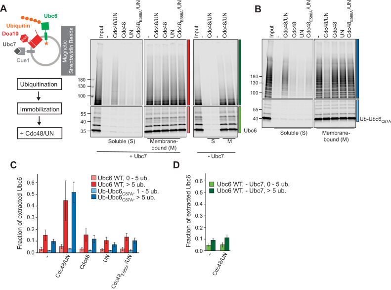
Figure 3. Cdc48-mediated Membrane Extraction of Ubc6.

(A) Extraction of Ubc6 by Cdc48 and Ufd1/Npl4 (UN). After ubiquitination, liposomes were immobilized (Figure 3—figure supplement 1,A to D). One bead equivalent was removed, and bound protein was eluted with SDS sample buffer (Input). Beads were then incubated with the indicated components. Soluble (S) and membrane-bound (M) material were analyzed by SDS-PAGE and fluorescence scanning. Colored bars indicate categorization of ubiquitin chain length as used for quantification in (C) and (D). For better visibility, bottom and top gel parts are scaled differently. See Figure 3—figure supplement 1E for uncut image. Final concentrations: 50 nM Ubc6, 20 nM Doa10, 0.1 µM Cdc48 hexamer, 0.1 µM Ufd1 and Npl4. (B) As in (A), but with Ub-Ubc6C87A instead of Ubc6. See Figure 3—figure supplement 1F for uncut image. (C) Quantification (mean ± SD) of three experiments as in (A) and (B). Ubiquitinated species were categorized according to ubiquitin chain length, as indicated in (A) and (B). The signal in the soluble fraction was normalized to that in the input. (D) Quantification (mean ± SD) of three experiments as in (A), when ubiquitination was performed in the absence of Ubc7.

Figure 3—source data 1. This file contains the quantification of the fraction of extracted Ubc6 from three experiments as in Figure 3A,B, as shown Figure 3C,D.
It also contains the quantification of the immobilization efficiency of Ubc6 (Figure 3—figure supplement 1C) and of Rhodamine-labeled lipids (Figure 3—figure supplement 1D), as well as the quantification of the extraction efficiency of Ubc6 modified with different ubiquitin chain lengths (Figure 3—figure supplement 1G).

Figure 3.

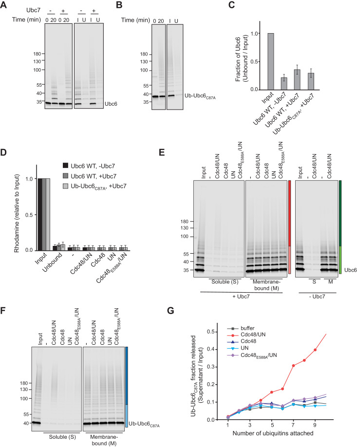
Figure 3—figure supplement 1. Cdc48-mediated extraction.

Figure 3—figure supplement 1.

(A) Samples from ubiquitination and immobilization under conditions described in Figure 3A. Liposomes containing Ubc6, Doa10 and Cue1 were incubated with E1, ubiquitin, and ATP, with or without Ubc7. After 20 min, the ubiquitination reaction was stopped by adding EDTA. Liposomes were then immobilized to streptavidin magnetic beads. Input (I) and unbound (U) fractions were analyzed by SDS-PAGE and fluorescence scanning. (B) Samples from ubiquitination and immobilization under conditions described in Figure 3B. As in (A), but with Ub-Ubc6C87A instead of Ubc6 and ubiquitination in presence of Ubc7. (C) Quantification (mean ± SD) of protein immobilization efficiency from three experiments as in (A) and (B). (D) Quantification (mean ± SD) of the efficiency of liposome immobilization in experiments described in Figure 3A,B, measuring the fluorescence of Rhodamine-PE that was co-reconstituted into liposomes. Rhodamine content of Input and Unbound fraction from immobilization reaction (as in (A) and (B)) as well as of the soluble fraction after extraction (as in Figure 3A,B) was determined. n = 3 independent experiments. (E) Uncut image with uniform scaling of Figure 3A. (F) Uncut image with uniform scaling of Figure 3B. (G) Comparison of extraction efficiency of Ub-Ubc6C87A modified with ubiquitin chains of different length as shown in Figure 3B.