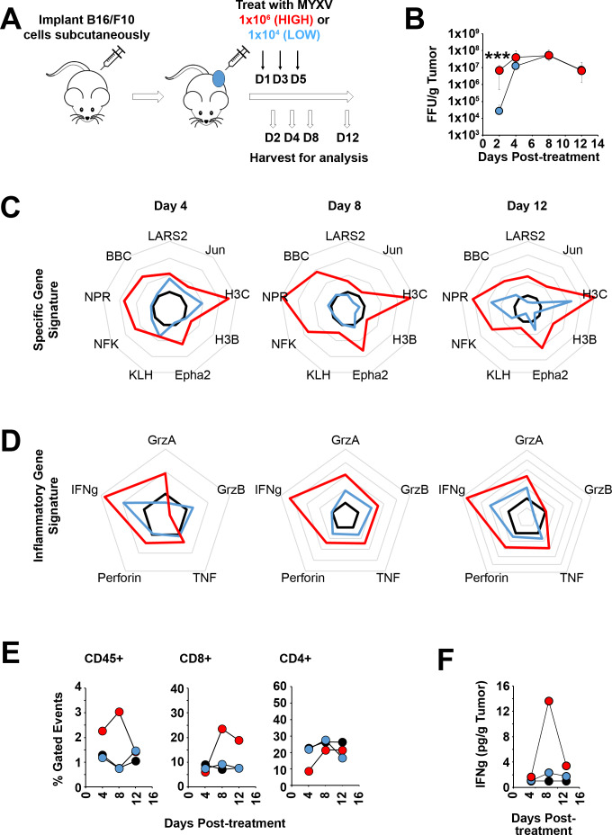
Figure 5.
Initial oncolytic dose determines a durable immunological program. (A) Schematic diagram of experimental design. C57/B6 mice were injected subcutaneously with B16/F10 cells. Once tumors reached ~25 mm2, animals were either mock treated or treated with three intratumorous injections of either a high (1×10⁶ foci forming units (FFU)) or low (1×10⁴ FFU) oncolytic dose of myxoma virus (MYXV). Tumors were then harvested 2, 4, 8, or 12 days after the initiation of treatment (n=5/time point/dose). (B) Quantitation of infectious virus in each tumor at each time point. Data are normalized to tumor mass and displayed as FFU/gram tissue. (C) Expression of the top 10 most significantly altered genes 4, 8, and 12 days post-treatment measured by quantitative-polylmerase chain reaction (qt-PCR). (D) Expression of a curated gene set made up of known adaptive immune mediators 4, 8, and 12 days post-treatment measured by qt-PCR. (E) Abundance of total CD45+ cells, CD8+ T cells, or CD4+ T cells within tumors treated as indicated at 4, 8, or 12 days post-treatment measured by flow cytometry. (E) Abundance of the T-cell effector molecule interferon-γ within tumors treated as indicated at 4, 8, or 12 days post-treatment measured by ELISA. Statistical significance was determined using unpaired Student’s t-test (***p<0.001). IFNg, interferon gamma.

