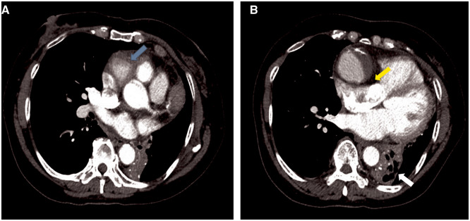
Figure 2.
Contrast-enhanced computed tomography of the chest, during the arterial phase in an axial orientation, depicting proximal aneurysmal dilatation of the right coronary artery (A, blue arrow) with evidence of right atrial compression (B, yellow arrow). There is an area of atelectasis within the left lower lobe with air bronchograms, with associated collapse of the left lower lobe (B, white arrow).

