Figure 3.

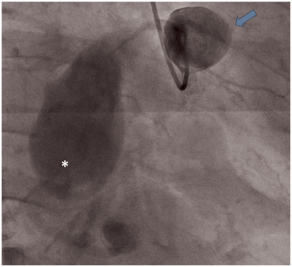
Invasive coronary angiography depicting giant saccular proximal (arrow) and giant fusiform mid (asterisk) right coronary artery aneurysm. Distal vessel segment is not well-opacified.

Invasive coronary angiography depicting giant saccular proximal (arrow) and giant fusiform mid (asterisk) right coronary artery aneurysm. Distal vessel segment is not well-opacified.