Figure 5.
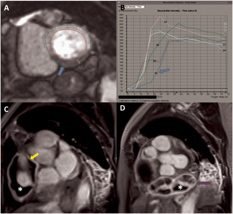
Gadolinium-enhanced cardiac magnetic resonance depicting resting hypoperfusion of the inferior wall on first-pass perfusion imaging (A), with reduced time to peak myocardial intensity on time signal analysis (B). Late gadolinium-enhanced cardiac magnetic resonance showing giant aneurysm in the mid right coronary artery with evidence of hyperenhancement of the aneurysm wall (yellow arrow) indicative of inflammation, with intraluminal thrombosis (asterisk) (C). Distal coronary artery giant aneurysm previously not visualized on coronary angiography with wall hyperenhancement (purple arrow) and thrombosis (asterisk) (D).

