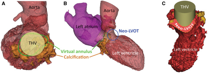
Figure 2.
Multi-detector computed tomography-derived 3D virtual model of the left heart with a prosthetic heart valve implanted. (A) Same model as in Figure 1D with a cylinder positioned in the mitral annulus representing the dimensions of a 26 mm Sapien3 transcatheter heart valve. (B) Overview of the left heart with the transcatheter heart valve implanted in mitral annulus, note the protrusion into the left ventricular outflow tract forming a neo-left ventricular outflow tract (blue circle). (C) Automatically calculated, the minimal neo-left ventricular outflow tract area in late-systolic phase with an implanted transcatheter heart valve 60% in the left atrium, 40% in the left ventricle was 212 mm2 (30% of original left ventricular outflow tract blocked).

