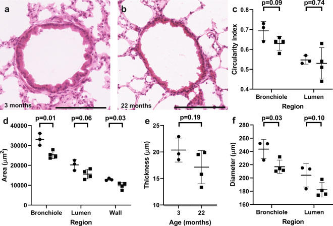
Figure 2.
Bronchioles become smaller in their cross-sectional area and diameter with age. Representative 40x images of H&E-stained distal lung bronchioles from (a) 3 month old and (b) 22 month old mice. Scale bars are 150 µm. Dotplots depicting (c) Circularity of the whole bronchiole and the lumen, (d) Area of the whole bronchiole, the bronchiolar lumen and the bronchiolar wall, (e) Thickness of the bronchiolar wall, (f) Diameter of the whole bronchiole and bronchiolar lumen. Circles represent 3 month old mice, and squares represent 22 month old mice. >7 images were analysed per mouse. Error bars are standard deviations. P-values refer to two-tailed T-test results.

