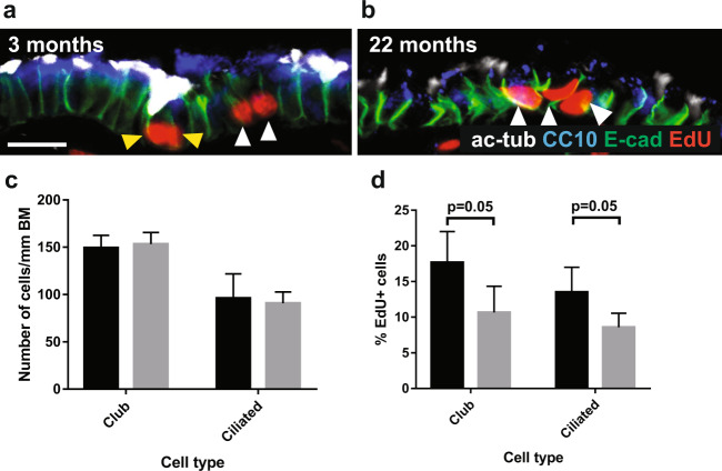
Figure 4.
Density of bronchiolar club and ciliated cells is maintained with aging, but proliferation rates of club cells decrease. 40x images of bronchioles from (a) 3 month old (n = 4) and (b) 22 month old (n = 4) mice treated with EdU ad libitum in drinking water for 14 days, stained for CC10 (blue), acetylated tubulin (white, ciliated cells), E-cadherin (green, epithelial cell adherence junctions), EdU (red, cells that have proliferated). Scale bars are 30 µm. Yellow arrowheads indicate EdU+ ciliated cells, white arrowheads indicate EdU+ club cells. Histograms depicting (c) density of club and ciliated cells per mm bronchiolar basement membrane, and (d) Percentage of club and ciliated cells that have incorporated EdU, indicating that they have undergone proliferation. >200 cells were counted per mouse, and 4 mice were in each age group. Black bars represent 3 month old mice, and grey bars represent 22 month old mice. Error bars are standard deviations, and p-values refer to two-tailed T-test results.

