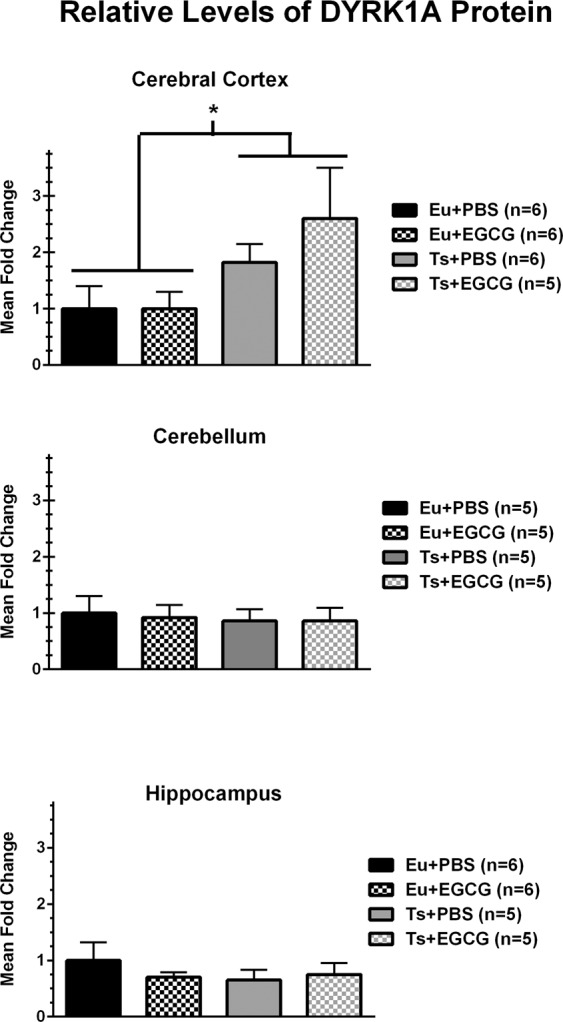
Figure 6.

Western blot analyses of relative DYRK1A protein levels for cerebral cortex, hippocampus, and cerebellum in 68-day-old euploid and trisomic male mice. DYRK1A from each brain region was normalized to actin as a loading control, then each sample ratio was expressed as a relative proportion of the mean value of the euploid-PBS control group for that brain region. DYRK1A was significantly overexpressed in trisomic mice only in the cerebral cortex (main effect of genotype, p = 0.034).
