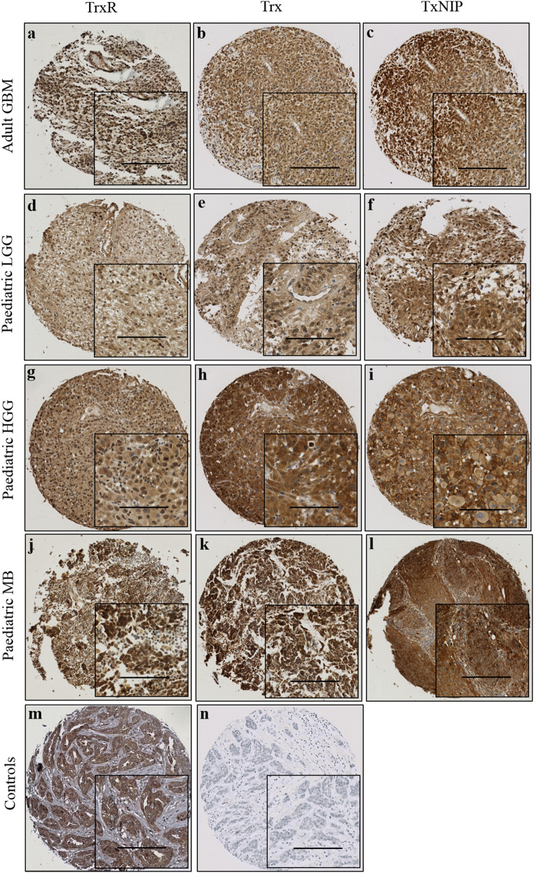
Fig. 1.
Representative photomicrographs of Trx system expression in different brain tumour types. Examples of TrxR (left panel), Trx (middle panel) and TxNIP (right panel) staining in a–c adult glioblastoma; d–f paediatric low-grade glioma; g–i paediatric high-grade glioma; j–l paediatric medulloblastoma; m representative positive control of TrxR on breast cancer tissue; n representative negative control with omission of the primary antibody on breast cancer tissue. Images were taken at × 10 magnification with × 20 magnification inset panel. Scale bar represents 100 μm

