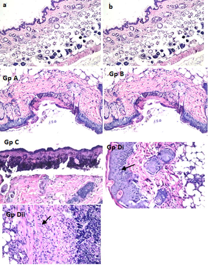
Figure 7.
Transverse stained (H&E) sections of the dorsal mice skin taken from different groups: (a) negative control, (b) irradiated control, GpA: animals were topically treated by Cur aqueous suspension, Gp B: animals were topically treated by Cur suspension, followed by irradiation after 1 h, Gp C: animals were topically treated by 100 mg of the selected Cur-loaded PEGylated lipid nanocarrier (PLN3), GpDi animals were topically treated by 100 mg of the selected Cur-loaded PEGylated lipid nanocarriers (PLN3), followed by irradiation after 1 h showing the acanthosis (increasing thickness of the epidermal layer, indicated by the arrow) and GpDii the subcutaneous tissues of the same group with focal inflammatory cells infiltration (indicated by the arrow).

