
Figure 3.
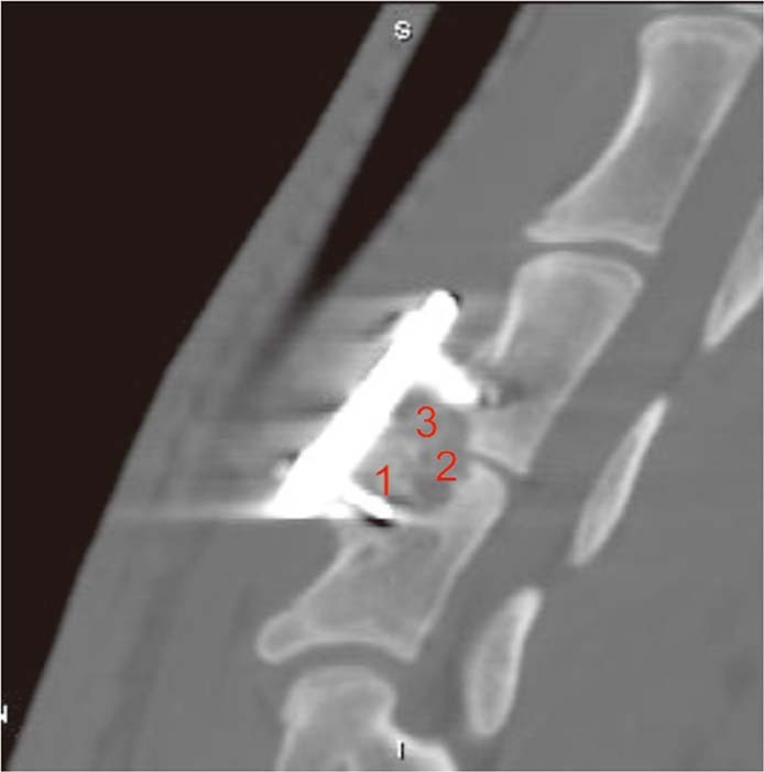
Radiographic assessment. Four sagittal planes at the C3 and C4 segments were reconstructed with 2-mm slice thickness. The interface between the implant and vertical bone on each plane was divided into 1, 2 and 3 zones for the degree of fusion status. The scoring criteria were described by van Dijk and graded as follows: 0= pseudoarthrosis; 1= ingrowth of bone with the strut securely fixed to vertebral bone above and below, but with a radiolucent discontinuity in the fusion mass; 2= arthrodesis with solid bone bridging the fusion area. The total points for each sample was 24 points.