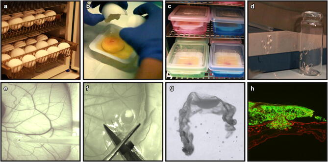
Figure 1.
Overview of the CAM-Delam assay. (a) Fertilized chick eggs were incubated horizontally. (b) On Day 3 of incubation, eggs were cracked and laid in a weighing boat in (c) an internal humidified chamber. (d) Silicon ring preparation. (e) On Day 10 of incubation, 1 × 10⁶ cells were seeded on the CAM using a pipette. (f) At different time points, the CAM membrane with the growing cancer cells were dissected out, fixed, sucrose treated and (g) positioned in frozen section medium. (h) Sectioned CAM with associated GFP+ cancer cells (green), followed by anti-Laminin immunohistochemistry (red).

