Figure 1.

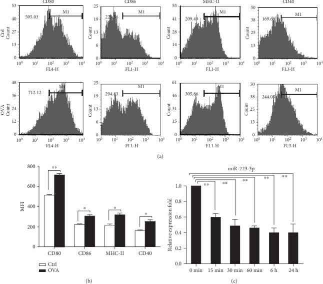
miR-223-3p was downregulated in OVA-induced DCs. (a) Mouse immature DCs were treated with 100 μg/ml OVA for 24 h and then stained with specific Abs against MHC-II, CD80, and CD86 for flow cytometry analysis. (b) The bar chart indicated the MFI of the DCs in each group. t-test, ∗P < 0.05, compared to the control group. (c) miR-223-3p expression in OVA-treated immature DCs. Immature DCs were incubated with OVA (100 μg/ml) for 24 h. Cells were collected at the indicated time points, and miR-223-3p expression was determined by RT-PCR. Data were shown as mean ± SD of three independent experiments. Post hoc Tukey's HSD test, ∗P < 0.05, ∗∗P < 0.01, compared to the 0 min control.
