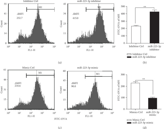
Figure 2.

miR-223-3p suppressed OVA endocytosis of BMDCs. Mouse immature DCs were transfected with the miR-223-3p inhibitor (a, b), miR-223-3p mimic (c, d), and the corresponding controls at a final concentration of 50 nM. After 24 h of transfection, DCs were incubated with FITC-OVA for 30 min and the endocytic activity (FITC-OVA uptake) of BMDCs was measured by flow cytometry. (b, d) The bar chart indicated the MFI in the gate of CD11c+ cells in each group. Similar results were obtained in three independent experiments. Data were shown as mean ± SD. t-test, ∗P < 0.05, ∗∗P < 0.01.
