
Fig. 1.
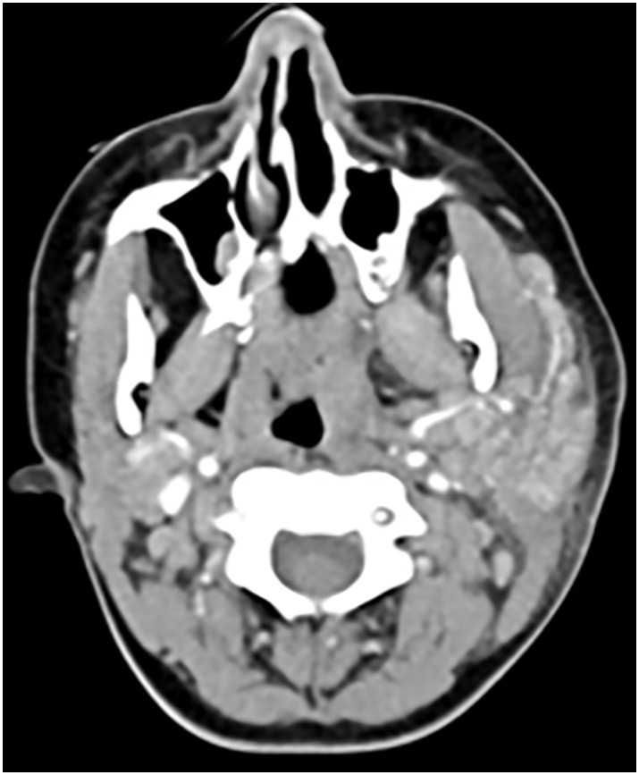
Axial image from a contrast enhanced CT of the neck demonstrates diffuse asymmetric swelling of the left parotid gland with surrounding fat stranding without associated obstructing stone, mass, or abscess.
Official websites use .gov
A
.gov website belongs to an official
government organization in the United States.
Secure .gov websites use HTTPS
A lock (
) or https:// means you've safely
connected to the .gov website. Share sensitive
information only on official, secure websites.
Axial image from a contrast enhanced CT of the neck demonstrates diffuse asymmetric swelling of the left parotid gland with surrounding fat stranding without associated obstructing stone, mass, or abscess.