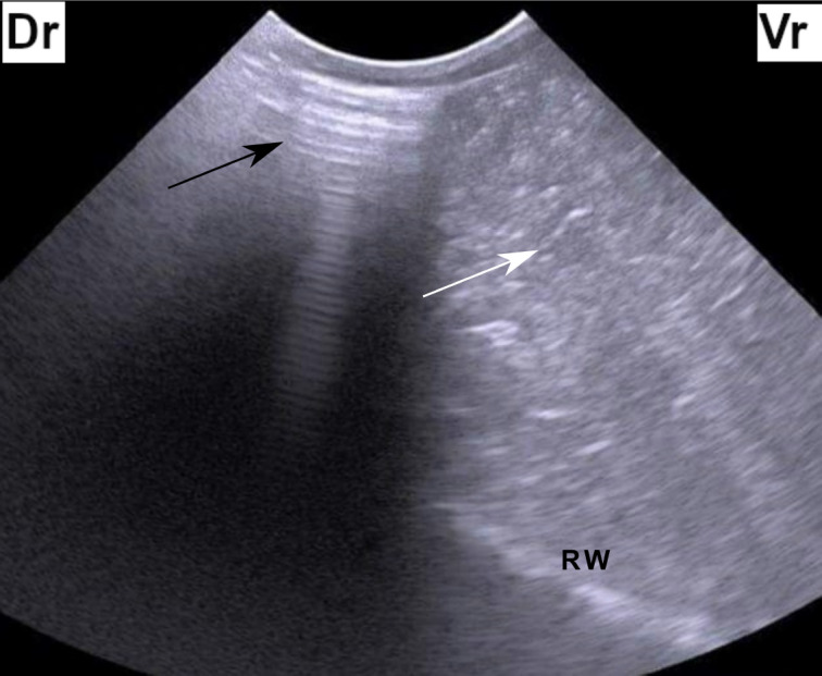
Figure 1. Ultrasonogram of the left abdomen at the level of the upper third of the 11th intercostal space in a cow identified with left-sided ping sounds. The figure shows reverberation artifact dorsally (black arrow) which represents a gas cap and hypoechoic fluid ventrally (white arrow). Dr = dorsal, Vr = Ventral, RW = Ruminal wall.

