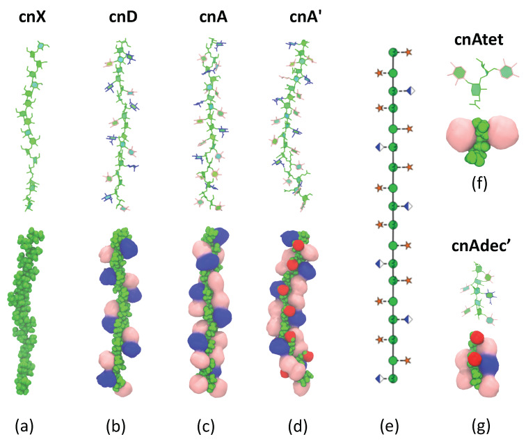
Figure 5.
Dominant conformations of the GXM molecules shown with two representations in the VDM package [26]—the PaperChain visualisation [27] to highlight the conformation of the backbone and space-filling representations to highlight the exposed binding surface. Representative conformations are shown for (a) the unsubstituted mannan backbone cnX; (b) cnD; (c) cnA; (d) and cnA’ (6-O-acetylated). A schematic for the arrangement of the side chain substitutions is shown in (e) using the SNFG symbols for the sugar residues [13,14]. Representative conformations of the chain fragments are shown for the tetrasaccahride (f) cnAtet and the 6-O-acetylated decasaccharide (g) cnAdec’. The chain substitutions in all representations are coloured as follows: DMan—green; DGlcA—blue; DXyl—pink and 6-OAc—red.

