
Figure 3.
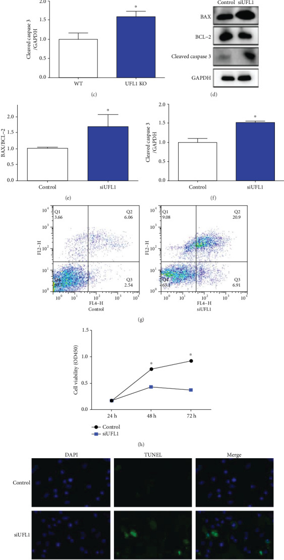
Effect UFL1 on proliferation and apoptosis in the mammary gland. (a–c) Mammary tissues were harvested from UFL1 KO mice after tamoxifen intraperitoneal treatment. (a) Samples were lysed, and the expressions of BAX, BCL and cleaved caspase 3 were analyzed by western blot. The relative intensity of (a) was plotted in (b) and (c). (d–i) HC11 cells were transfected with siUFL1 or control before being harvested. (d) Western blot analysis of process apoptosis-related proteins BAX, BCL, and cleaved caspase 3 levels. The relative intensity of (d) was plotted in (e) and (f). (g) HC11 cells transfected with UFL1 siRNA were stained with annexin and PI tested to flow cytometry followed and then quantification of apoptosis ratio. (h) Cell viability was detected by CCK-8 analysis. (I) TUNEL analysis was used for determining the apoptosis of HC11 cells followed by UFL1 siRNA treatment. TUNEL positive was shown as green fluorescence, and nuclei were stained with DAPI as blue fluorescence. Scale bar : 20 μm. Results were analyzed by means ± SEM. ∗ explores significant difference (P < 0.05). N.S. indicates no significant difference (P > 0.05).