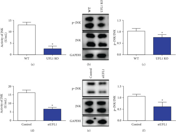
Figure 7.

UFL1 suppressed the activation of JNK. (a–c) Mammary gland tissues were collected from UFL1 KO mice after tamoxifen intraperitoneal treatment. (a) The activity of JNK was tested by ELISA using a JNK activity assay kit. (b, c) The abundances of p-JNK and JNK proteins from mammary tissues of UFL1 KO mice were analyzed by western blot determined. (d–f) Cells were transfected with control siRNA or UFL1 siRNA for 72 h. (d) After treatment as previously described, cells were lysed in PBS, and then the activity of JNK was expressed by the JNK activity assay kit. (e, f) The levels of p-JNK and JNK were detected by western blot. The relative intensity was p-JNK relative to the JNK level. Bars were expressed as the mean ± SEM of the data. ∗ indicates significant difference (P < 0.05).
