. 2020 Apr 20;7(2):94–106. doi: 10.3934/Neuroscience.2020007

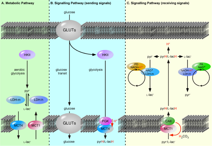
Figure 1. The Processing of Glucose as a Metabolic versus Signalling Pathways. (A) The metabolic pathway is understood as a solely cytosolic process, were hexokinase II (HKII) is set as first enzyme and initiates the gradual degradation of the carbon backbone of glucose to pyruvate (pyr). The extrapolation of so-called “aerobic glycolysis” to include lactate dehydrogenases (LDHs) and proton-linked monocarboxylate transporters (MCTs) encounters two pitfalls. First, the reactions are illustrated with arrows indicating an equilibrium reaction catalyzed by single enzyme and not as two independent and sterically separated metabolic pathways. Second, following a sequential pathway, i.e., that the product of enzyme A is substrate of enzyme B, results in MCT catalyzed transfer of l-lactate (l-lac), instead of monocarboxylic acids. (B, C) Glucose metabolism as signalling pathways can be divided in a “sending” pathway and a “receiving” pathway. The transferred signalling molecules are glucose, pyruvic acid (pyrH) and lactic acid (l-lacH). In the “sending” pathway, the MCT4·phosphoglycerate kinase (PGK) complex exports l-lacH and pyrH depending on the glycolysis rate or PGK activity. The “receiving” pathway utilizes the MCT1·carbonic anhydrase II (CAII) complex, which import monocarboxylic acids depending on cellular carbonic acid (H2CO3) flow. The imported l-lacH and pyrH are detected by heart lactate dehydrogenase (LDH-h) and muscle LDH (LDH-m) complexes, respectively. Here, LDH-m acts as a proton acceptor (PA) protein, whereas the LDH-h acts as proton donor (PD) protein and catalyzes the oxidation of l-lac forming the proton carrier, NADH-H+. The LDH-m complexes are considered part of the glycolytic pathway, detecting the metabolism of glucose. The separation of unidirectionally catalyzed reactions into distinct pathways provides an explanation for why pancreatic β-cells release insulin in response to imported pyrH, similar to glucose [15].

Figure 1.